41st Annual Diagnostic Imaging: On Cape Cod



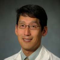
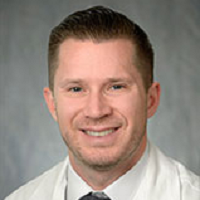
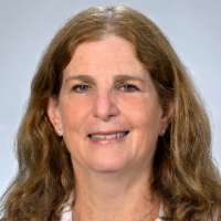
Speakers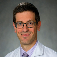
OVERVIEW
41st Annual Diagnostic Imaging: On Cape Cod is organized by Penn Medicine - Department of Radiology and will be held from Jul 08 - 12, 2019 at Chatham Bars Inn, Chatham, Massachusetts, United States of America.
Target Audience:
This CME activity is designed to educate diagnostic imaging physicians who perform and interpret imaging examinations.
CME Credits: 23.5
Physician Continuing Medical Education
Meeting:
The Postgraduate Institute for Medicine designates this live activity for a maximum of 23.5 AMA PRA Category 1 Credit(s)™. Physicians should claim only the credit commensurate with the extent of their participation in the activity.
The credit breakdown by day:
Day 1: 4.75
Day 2: 4.75
Day 3: 4.75
Day 4: 4.75
Day 5: 4.5
Workshop:
The Postgraduate Institute for Medicine designates this live activity for a maximum of 2 AMA PRA Category 1 Credit(s)™. Physicians should claim only the credit commensurate with the extent of their participation in the activity.
Course Overview:
Our annual five-day summer course, now in its 41st year, is designed for radiologists in clinical practice. This year's course will include lectures related to challenging cases and diagnostic pitfalls. Speakers will discuss advanced techniques as well as offer practical tips and share imaging protocols to enhance image acquisition and interpretation skills. Additionally, the speakers will emphasize indications for complimentary use of different imaging modalities. SAM credits will be offered throughout the week
Learning Objectives:
Upon completion of this activity, participants should be able to:
• Effectively utilize US in the evaluation of the thyroid nodules and diffuse thyroid disease
• Apply the updated guidelines for recommending fine-needle aspiration, molecular testing and follow up imaging of thyroid nodules
• Describe the specific characteristics of diffuse and focal pulmonary lesions
• Apply the current pathological and radiological classification of adenocarcinoma spectrum lesions
• Describe the CT and MR imaging of features of mediastinal lesions
• Describe the MR appearance of abnormalities in the brain, liver, pancreas and gastrointestinal track
• Utilize the LI-RADS reporting system of hepatocellular carcinoma
• Review implementation, interpretation of and appropriate indications for low dose CT in the abdomen and pelvis
• Describe the MR appearance of knee and spine pathology and injuries
• Describe the imaging of features of mediastinal lesions
• Utilize the WHO reporting systems for classifying brain neoplasms
• Identify post-operative complications in the spine
• Discuss methods to improve imaging of the post-operative spine
Focused Musculoskeletal Ultrasound Hands-On Workshop:
In this new 2-hour hands-on workshop, participants will practice scanning of common musculoskeletal ultrasound examinations. This workshop will cover ultrasound of major joints for conditions including effusion, bursitis and common tendon pathology. Simulation of ultrasound-guided injections for musculoskeletal pathology in the upper and lower extremity will be performed as well. This workshop is suitable for radiologists with little or no prior musculoskeletal ultrasound experience. Limited enrollment. Participants will select from two 2-hour time slots during registration.
Learning Objectives:
At the conclusion of this session, participants should:
• Describe the scanning technique for common musculoskeletal ultrasound examinations
• Be able to perform basic musculoskeletal ultrasound examinations in the upper and lower extremity
KEY DATES

08 Jul, 2019
Event Start Date

12 Jul, 2019
Event End Date
SPECIALITIES
RadiologyRadiological SciencesMusculoskeletal RadiologyThoracic RadiologyAbdominal RadiologyNeuroradiologySUGGESTED HOTELS
CONFERENCE VENUE
 Chatham Bars Inn
RELATED CONFERENCES & LIVE WEBINARS
By American Medical Seminars (AMS),...
Feb 15 - 18, 2027 | Sarasota, Florida,...
Radiology
20 AMA PRA Category 1 Credits™
...
...

By Provider Practice Essentials...
Mar 19 - 20, 2027 | Dallas, Texas, USA
State required course for Illinois ...
23 CME/CE
Quick Links