Introduction to Carotid & Peripheral Vascular Duplex/color Flow Imaging - Virtual Conference



Speakers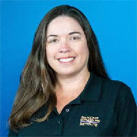



OVERVIEW
Introduction to Carotid & Peripheral Vascular Duplex/color Flow Imaging - Virtual Conference is organized by Gulfcoast Ultrasound Institute (GCUS) Inc and will be held from Sep 27 - 30, 2021.
CME Accreditation
The Gulfcoast Ultrasound Institute designates this internet live activity for a maximum of 8.00 AMA PRA Category 1 Credit(s)™. Physicians should claim only the credit commensurate with the extent of their participation in the activity.
Description
Introduction to Carotid & Peripheral Vascular Duplex/Color Flow Imaging Virtual Conference is taught by leading vascular ultrasound experts, and is specifically designed for physicians, sonographers and other medical professionals who need Carotid & Peripheral Vascular Duplex/Color Flow Imaging training. These four day Carotid & Peripheral Vascular Duplex/Color Flow Imaging ultrasound courses includes 130+ interactive case presentations. In this (4) four day long vascular program, participants will learn ultrasound techniques for the entire vascular system.
Topics:
• Doppler & Color Fundamentals
• Carotid Anatomy & Physiology
• Normal Spectral Analysis
• Carotid Scan Protocol
• Disease Progression & Stenosis Assessment
• Carotid Imaging Pitfalls
• Interactive Case Studies
• Evaluation & Measurement of Intima-Media Thickness
• Carotid Abnormalities
• How to Structure a Report
• Venous Anatomy, Scan Techniques & Normal Characteristics
• Duplex / Color Evaluation of LE, UE DVT
• Evaluation of Venous Insufficiency
• Duplex / Color Evaluation for Venous Ablation Procedures
• Types of Vein Ablation Procedures
• Pre-Procedure Mapping
• Post-procedure Mapping
• LE Arterial Anatomy & Physiology
• Clinical Exam & Indirect Testing
• Direct Testing & Duplex Scanning
• Live Demo: Arterial Duplex of Lower Arterial Disease
Objectives:
• Increase the participant’s knowledge to better perform and/or interpret Carotid Duplex/Color Flow Imaging ultrasound examinations.
• Apply knowledge of the anatomy/physiology of the cerebrovascular system into the Carotid Duplex examination.
• Cite Doppler/color physics principles and be able to (sonographers) apply these principles to optimize system controls or (physicians) utilize this information for identifying technical errors which may result in misdiagnosis.
• Perform routine scan protocols and Doppler calculations in a complete carotid duplex/color examination.
• Differentiate normal/abnormal spectral Doppler/color characteristics.
• List methods for obtaining quantitative information and state the diagnostic relevance of each measurement.
• Characterize plaque morphology and other pathology associated with cerebral vascular disease.
• Perform Intima-Media Thickness measurements and state the clinical significance as a screening method for cardiovascular disease.
• Integrate the information to include and prepare a structured report for a carotid ultrasound examination.
• Apply diagnostic criteria for accurate interpretation of carotid duplex/color flow examinations.
• Increase the participant’s knowledge to better perform and/or interpret upper and lower Peripheral Vascular ultrasound examinations.
• Apply knowledge of the anatomy/physiology of the upper and lower extremity venous and arterial systems into the venous and arterial duplex and physiologic testing examinations.
• Cite Doppler/color physics and be able to (sonographers) apply these principles to optimize system controls and/or (physicians) utilize this information for recognizing technical errors which may result in misdiagnosis.
• Perform routine scan protocols, and document Doppler waveforms for lower extremity arterial and venous evaluations of the upper and lower extremity.
• Differentiate normal/abnormal imaging, spectral Doppler and color characteristics for identifying arterial and venous disease.
• State the indications and applications of indirect testing methods for lower arterial disease.
• Demonstrate vein mapping techniques to identify suitability as a potential arterial bypass graft.
• Perform routine scan protocols and document Doppler waveforms for venous evaluation of the lower extremities, including pre and post vein ablation evaluation.
• State the role of ultrasound in the diagnosis and treatment of venous insufficiency.
• Perform evaluation for venous insufficiency and patency of perforators for vein therapy treatment.
KEY DATES

27 Sep, 2021
Event Start Date

30 Sep, 2021
Event End Date
ACCREDITATION & CREDITS

TARGET AUDIENCE
PhysicianPhysician AssistantSonographerPrimary Care PhysicianCardiologistsSurgeonsRadiologistsVascular SurgeonNeurologistsInternal MedicineSPECIALITIES
CardiologyRadiologyNeurologyVascular SurgerySurgeryPrimary CareSonographyPhysician AssistantsRELATED CONFERENCES & LIVE WEBINARS

Quick Links